Friday, December 24, 2010

Nature's Stained Glass Windows


Looking like a stained glass window, this is the remains of the seed pod of one of last summer's garden flowers, Lunaria annua (commonly known as honesty, because the dry seed pods resemble silver coins), magnified around two hundred times and viewed using polarised light.


When honesty seed pods ripen they are flattened and composed of three components. Imagine three large 'coins' joined to each other all around their rims, with the central 'coin' attached to the plant via a stalk. Swelling seeds are attached to the rim of both faces of the central 'coin' via their own slender stalks, visible when sunlight shines through the whole structure.

When the pod dries out and ripens the tensions in the drying, contracting cells of the walls of the 'coins' tear them apart around their rims, so the two outer 'coins' detach and flutter away in the breeze, followed by the winged seeds, leaving the central 'coin' attached to the dead plant and surviving deep into winter.

These are the cells of that central, surviving 'coin' magnified about one hundred times and using ordinary white light....


... and these are the dazzling interference colours generated when polarised light is used.


At two hundred times magnification it's clear that the 'coin' is formed from two layers of cells, orientated at different angles, so that the tensions they develop when they dry will twist and distort the 'coin' and help to rip apart the sutures with the outer 'coins'.


 At four hundred times magnification you can clearly see the pores through the thick cell walls which were the plasmodesmata - the channels of communication between the cytoplasmic contents of one cell and the next, while the whole structure was still alive and the seeds were still developing.




The dry, dead cells form intricate patterns...


... whose colours change as they are rotated in the plane of the polarised light.

Saturday, December 18, 2010

The Colour Purple

This rather beautiful flower is Tibouchina urvilleana and the purple of its petals is due to the presence of anthocyanin pigments, which are dissolved in the cytoplasm of the petal cells.


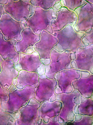
If you magnify the petal surface about 200 times you can see the way in which the petal cells fit together, like pieces of a jigsaw puzzle. This piece of petal is mounted in water but if it's transferred to a concentrated solution of sugar ...


  ... the water in the cell begins to flow out through the semi-permeable cell membrane by the process of osmosis, with water travelling out from the less concentrated solution in the cell to the more concentrated solution surrounding it. Within a few minutes spaces become visible between the cytoplasm and the cell wall, where the cytoplasm shrinks ....


... and within a few more minutes the cytoplasmic contents of the cell have shrunk even further, so the purple anthocyanin pigment becomes even more concentrated in the remaining cytoplasm.


  While I was looking at the petals I noticed something unusual around their edge  - a fringe of microscopic hairs, invisible to the naked eye.


 Each hair is tipped with a  glandular head.


... that looks as through it may contain oils.

What are these hairs for? Secreting aromatic compounds that attract insect pollinators, perhaps?

Friday, December 3, 2010

Sticky Jack

This is a cross section of the stem of the plant commonly known as goosegrass or sticky Jack and more scientifically as Galium aparine. Sticky Jack is a very common weed that scrambles up through other vegetation using its covering of hooked hairs on the stem and leaves and which sticks to clothing with these when kids throw handfuls of the stuff at each other.
This image was produced using fluorescence microscopy, staining the cells with compounds that bind to the cell walls and fluoresce. The blue cells have walls made of cellulose and their blue fluorescence is due to the calcofluor that they've been stained with, which fluoresces blue in ultraviolet light. Calcofluor has been used as a 'blue whitener' in washing powders - it binds to the cellulose in cotton fabrics and fluoresces faintly blue in the UV component of sunlight. The yellow staining is due to another fluorescent dye (fluorochrome) called auramine O, which binds to cutin in the outer cuticle of the plant, and to dead, lignified cell walls that give the stem its strength - and it fluoresces yellow. The cuticle in this cross section is the thin yellow line covering the outer surface of the section. The yellow circle in the centre is composed of dead, lignified cells - not particularly well developed in goosegrass because it scrambles over surrounding vegetation rather then investing resources in producing a stout lignified stem of its own.

Saturday, November 27, 2010

Strings Attached

Deciding on the prime time to pick a bean pod is s tricky business. Leave it too late and the pod will become tough and stringy - and the reason for that is because as it grows the pod begins to prepare to shed its seeds. Members of the pea family - the leguminosae - carry their seeds in pods that naturally become brittle when they dry and ripen, when tensions developed along the suture between the two pod halves and in the pod wall eventually become so great that the pod splits open violently, hurling out the seeds. Plant breeders have worked hard to breed this trait out of legume crops, but species like runner bean still produce long strands of woody, lignified cells in their pod walls as they ripen. In this fluorescence micrograph, showing a cross section of the upper suture of a developing pod, the bright yellow arcs of cells at the top are the 'strings' that you need to strip out of the pod before you eat and cook it if you've left it too long before harvest. The yellow cells just creeping into the picture at bottom left belong to the parchment layer that develops in the pod wall. Together, these thock-walled cells develop the tensions in the pod as it dries that will eventually split it open along its longitudinal sutures and release the seeds.

Monday, November 22, 2010

Twister

To appreciate the true beauty of mosses you really need to explore them with a hand lens or low power microscope. This is Tortula muralis, wall screw-moss and to find out how it acquired that colloquial name you need to take a close look at the spore capsules.

Wall screw moss grows in the mortar-filled cracks in walls, where it produces spore capsules that are carried aloft on stalks that are a couple of centimetres long at maturity. These are capsules in the very early stages of development, before their stalks lengthen, but if you take a really close look at a mature spore capsule....

 ...it looks like this. Notice how the capsule's stalk (seta) has twisted helically. If you take a close look at the capsule (double click on the image for an enlarged version) you can see that most of it is sheathed in a membranous covering - the calyptra. Gently pulling this off with a pair of forceps reveals....
 ... a lidded capsule underneath and if you pull the lid (operculum) away....


... it reveals a screw thread-like arrangement (the peristome) underneath, that gives the moss its common name. These threads twist up tightly in moist air but untwist in a dry atmosphere, allowing the minute spores to be shaken out when the seta trembles in the wind. In this image you can see that the operculum that has been removed has become temporarily stuck to the base of the capsule - normally it will just fall away.

Thursday, November 11, 2010

Control Centre

This is a cell from a developing seed cotyledon of a broad bean Vicia faba plant. In the centre you can see the nucleus, floating like an icy-blue planet in a universe of cytoplasm - mission control, home the the DNA molecules that encode all the instructions for making the cell and, indeed, the whole plant. I stained the cell with a fluorescent dye that binds to the DNA in the nucleus and fluoresces blue when it's illuminated with ultraviolet light - the DNA (chromatin) is visible as the bright blue flecks on the nuclear membrane. The large, brightly fluorescing spot inside the nucleus is the nucleolus, where DNA is transcribed into ribosomal RNA subunits that are transported out of the nucleus and assembled into ribosomes in the cytoplasm. There the ribosomes form part of the machinery that translates the genetic code carried by messenger RNA, which is transcribed from the DNA in the nucleus, into proteins that are assembled from amino acid subunits. The nucleolus is highly visible in this cell because cotyledon cells in the seed manufacture and store proteins that are used when the seed germinates, to support the early growth of the seedling; this is a very busy nucleus and nucleolus because at this stage the cell is making a lot of protein. During early cotyledon development the cells are loosely packed together and you can see large triangular intercellular spaces between them. Later these will disappear, as the cells become packed full of proteins, lipids and starch, the cell walls thicken and the seed dries out during the seed ripening process. N.B. The cell walls are fluorescing blue in this image because of their own inherent biophysical properties, not because they contain DNA like that which is fluorescing blue in the nucleus.

Tuesday, November 2, 2010

Trees: the Inside Story


Almost as soon as plants colonised the land surface they began to compete for light, struggling to grow out of each other’s mutual shade. The ultimate solution, adopted by trees,  was to produce woody stems and grow tall, shading out competitors below. It's a very successful strategy - left to their own devices, many terrestrial ecosystems where water and warmth are adequate become forests. These (above) are cross sections of stems of two sycamore Acer pseudoplatanus seedlings, just a couple of weeks after germinating from a seed in spring, and already they have begun to produce woody thickening in some of their cells, visible here as the bright yellow fluorescent staining inside the stem (on the periphery of the large pith cells in its core). The very narrow yellow fluorescent line around the perimeter of the stem is the waxy cuticle secreted by the epidermal cells that protects the young stem – just a couple of millimetres in diameter at this stage - from water loss and invasion by pathogens. Double-click on the image for a clearer picture.

Fast-forward almost three years now and this seedling has grown into a sapling. In this cross section of a three year old lime (Tilia sp.) stem the big cells at the core are the pith. The three concentric rings of brown cells outside of that contain the xylem vessels that conduct water up and down the stem. They’re dead and their walls are strengthened with woody lignin, producing a strong, rigid support for the fast growing shoot and leaves. The width of those annual rings varies according the growing season – but I suspect that the outer, most recent ring is narrower because this shoot was harvested for microscopic sectioning sometime in mid-summer, before that year's annual growth was complete. Take a close look at the outer edge of the outer annual ring of xylem (double click the image to enlarge) and you may just be able to make out a distinct narrow zone of very small blue-stained cells, just a  few cells thick (at about 7 o'clock on the section). This is the cambium – the thin layer of living cells that divides to produce dead xylem cells on its inner face and living phloem cells, that conduct sugars from the leaves to the rest of the plant, on the outer side. Together the phloem and cambium are only a few cells thick and represent the most important living tissue inside the tree. Their protection is vital for the tree’s survival, so they are covered by a thick layer of bark tissue, also stained blue where the cells are alive but showing as grey-brown on the outer surface of the twig, where they are dying or dead. This is the tree’s waterproof,  self-repairing, insulating,  wound healing tissue, protecting the delicate living layer of cells inside. Growing tall by producing annual rings of growth is a long-term investment for a plant which only reaches full size after decade of growth, but the return on investment can then continue over centuries – and in some cases millennia - of annual flowering and seed production. As the stem adds annual rings, expanding in girth with every succeeding year, the outer dead bark layer splits into characteristic patterns, depending on the tree species.  The line of red cells in the bark tissues are fibres - dead cells that strengthen the young stem.

Monday, October 18, 2010

More Smut - Sex-change in Campion Flowers

A while back I posted some pictures of the smut fungus Microbotryum violaceum infecting the anthers of white campion .The flower above is a pink campion - probably a hybrid between white campion Silene latifolia and red campion Silene dioica - whose stamens are also full of the brown fungal spores. Both red and white campions exist as separate male and female plants. This fungus fills the anthers of male plants with its spores which are carried from flower to flower by pollinating insects, but it also brings about a change in the sex of female flowers, so that the female structures are suppressed and appendages called staminodes develop into stamen-like structures that produce the fungal spores and attract insects that are fooled into thinking they are collecting pollen grains. So, when the whole local population has been infected with the sex-changing smut fungus, how do you separate the genuine males from the gender switching females? 

 
 You need to look closely, under the microscope. If the 'stamens' are simply full of fingal spores, like those above, then you are looking a gender-switching female, but.....

 
......  if there are a few large pollen grains amongst the tiny fungal spores, then you are looking at a genuine male plant that was infected after the pollen grains began to develop.

Thursday, September 23, 2010

Harvestman

I found this delightful little harvestman amongst some raspberries that I picked yesterday evening. Although they are carnivores, they do seem to have a liking for ripe fruit.
Despite their spider-like appearance and the fact that they belong to the Arachnids, harvestmen are not true spiders but belong to an order of their own known as the Opiliones, distingished from spiders by their globular bodies, unlike those of true spiders which are divided into two parts - the separate thorax and abdomen, separated by a constriction. Harvestmen always seem to have an other-worldly appearance and, scaled up to monstrous proportions, wouldn't be out of place in a science fiction movie. Many of their sensory functions are located in their legs and if you watch the way in which they use these - particularly the second pair that are often far longer than the others - it's quite clear that they are using them to feel and taste their way around their habitat. Nevertheless, when danger threatens they can shed a leg (autotomy) and leave it twitching on the ground, to deflect the attention of  a predator. Unlike true spiders, they can't regrow limbs after moulting, so this desperate measure leaves them short of a limb and sensorily deprived. Before they resort to limb-shedding, they often exude an unpleasant-smelling liquid from their leg joints which you can smell if you hold an irritated harvestman in your hand. This isn't an infallible deterrent - I've watched robins feeding their brood with harvestmen.


One of the most remarkable features of harvestmen is the arrangement of their eyes, in a kind of turret called an ocularium, high up on their back. This arrangement makes sense when you get down to a harvestman's eye-level ...

... when it's clear this this gives it 360 degree vision around and above its globular body....


..... although one can only guess at how much detail those unblinking black eyes resolve.

Nevertheless, whatever angle you approach from a harvestman always has it covered........one of those eyes always seems to have you in its sights...

There's an excellent little booklet called British Harvestmen by J.H.P. Sankey and T.H.Savory (Synopses of the British Fauna (New Series) No. 4  ISBN 0 12 619050) that not only provides fascinating detail about their biology but also contains some delightful little anecdotes. For example, some species apparently kill their prey by positioning their globular bodies over their victim that's imprisoned by their legs and then bouncing up and down on those long legs, 'pile-driving' the unfortunate prey. Others have been noted for a prediliction for marmalade sandwiches from a picnic and on one occasion ink from ink wells (although this one refused black ink and would only drink the red stuff). 

I haven't got around to identifying the species of this individual yet and if there's anyone out there who can help me out, I'd be grateful....

Monday, September 13, 2010

Moth Pointillist Colour Patterns


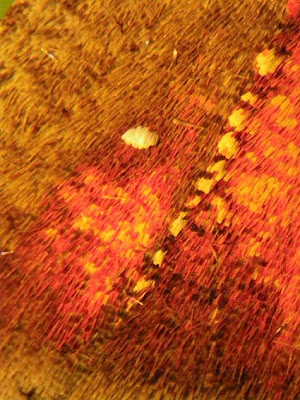
I found this herald moth Scoliopteryx libatrix, with these beautiful orange markings on its wings, in my garden.


The whole moth is covered with scales, of various shapes, sizes and colours over its whole body, even to the extent that its legs are clothed in this rather fetching pattern of alternating black and white rings.


The main body is covered with fine hair-like scales, but the scales on the wings are....


... much broader, although they vary in width and colour. One interesting feature is that the patches of colour that look fiery orange to the naked eye are composed of a mixture of pinkish-red scales interspersed with variable numbers of yellow scales. The whole effect is reminiscent of colours produced in Pointillist paintings, of the kind made famous by George Seurat. By Seurat's day (1859-1891) the study of colour had revealed that the close juxtaposition of points of two colours could produce the effect of a third colour when viewed from a distance and Seurat exploited this in his meticulously executed paintings. The computer monitor screen that you are viewing this blogpost on uses a similar principle, of coloured dots, to produce its vast range of colours. Butterflies and moths have been exploiting the same phenomenon for millions of years, to either make themselves conspicuous to mates or generate camouflage patterns.
The herald moth's wings also carry small clusters of distinctive white scales, like those just above the 'orange' patch here, which I suspect may be scent scales that emit pheromones recognised by other individuals of the same species.

Monday, September 6, 2010

Brittle Stars

The swaying fronds of red seaweed that fringe rockpools near the low tide level on the seashore are home to a wealth of miniature marine life, less spectacular than the inhabitants of coral reefs but every bit as intriguing. I found scores of these tiny brittle stars, the largest no larger than a centimetre across (including arms), on a visit to the Northumberland coast at the weekend. Brittle stars, or ophiuroids, are relatives of starfish and sea urchins, in the phylum Echinodermata (which means spiny skin - a feature many members of the phylum share). The view above is of the underside of one of the brttle stars, showing the mouth fringed with five teeth formed from calcareous plates.

Seen from above, five arms radiate from the pentangular body. Each arm is formed from articulated segments linked by muscles and these are very flexible, so the animal often curls the tip of an arm around a seaweed frond to stop itself from being washed away by currents. If it's alarmed the muscles between the arm segments contract and then the arms become very brittle.....


... and it doesn't take much force to snap them, as has happened here with the upper arm. This is not a problem, as....

... arms can easily be regenerated, as is happening here with the middle, lower arm. This capacity for shedding and regenerating arms is analogous to the way that lizards shed their tails (autotomy) if they are picked up by that appendage.

At higher magnification you can see the anatomy of the arms more clearly. Each calcareous segment bears spines and a pair of tube feet, that are all interconnected by a hydaulic system of radial canals that run along the arms and a ring canal that runs around the central body. Local relaxation or contraction of muscles, compressing liquid within, elongates or retracts the tube feet.
Unlike the tube feet of starfish which have suckers on their tips and are used for 'walking', those of brittle stars are primarily for sensory purposes and to assist in feeding, by secreting adhesive mucus. In this higher magnification image you can see that the tube foot is hollow.

The ring of tube feet around the mouth on the underside, where the arms converge, help to sweep food particles beyond the five calcareous teeth)....
 

.... into the muscular oesophagous, and then into ....

... the stomach. The tiny central body also contains gonads, that produce eggs and sperm that give rise to the planktonic ophiopluteus larvae.

When they're fully grown some brittle star species can reach 60 centimetres in diameter (not in Britain, though), but they all begin life as planktonic larvae, often settling into the shelter of seaweeds on the nursery slopes of rock pools or coastal shallow seas, which are of such importance for the health of the oceans.

You can see a YouTube vieo sequence of an adult brittlestar here.


Thursday, September 2, 2010

Plant Plumbing


Swiss cheese plant Monstera deliciosa is commonly grown as a decorative house plant but in its native Mexican rainforests it's a rampant climber, using its adventitious roots to cling to trees and climbing in much the same manner as ivy in temperate woodlands. Those holes in the Monstera leaf, whose resemblance to holes in Swiss cheese account for its name, let flecks of sunlight filter through to the layers of leaves below, all of which are transpiring water from their surface. If you cut a section through the stem, you can see the internal pipework that conducts water from the roots to the leaves.


In this transverse section of adventitious root, stained with fluorescent dyes that colour dead, woody cell walls yellow and living cellulose cell walls blue you can see the various cells that conduct liquids up and down the root. Embedded in that thick-walled strengthening tissue that gives the root (which in this case is used for clasping tree trunks and branches - this plant is a tropical climber)  rigidity and are fluorescing yellow, are large vessels that conduct water in a continuous tensile column from the roots to the leaf, pulled upwards by evaporation from the leaf surface. The smaller tubes, lined with a layer of blue-fluorescing cell walls, may be resin ducts. The outer cells on the left, part of the ring of small bundles of living cells that encircle the root, are the phloem cells that conduct sugars manufactured by photosynthesis in the leaf to other parts of the plant. Swiss cheese plants are such familiar items of interior decor that they hardly attract a second glance, but they have extraordinary hidden beauty, only visible under the microscope