Wednesday, October 24, 2012

Mosquito larvae...


Our unusually wet summer has provided plenty of opportunities for breeding mosquitoes, with water butts and containers retaining pools of water all through the year. The water butt attached to our greenhouse has been swarming with culicine mosquito larvae.


The larvae hang from the surface film, breathing through a  siphon tube, which you can see in this image on the tail of the larva; you can see a small bubble of air attached to the siphon tip. The tiniest disturbance of the surface film sends the larvae wriggling down into the depths.


For images of an adult culicine mosquito, click here and here.


Thursday, August 30, 2012

How to Recruit an Army


Plants primarily secrete nectar as an energy source to tempt pollinators to visit their flowers, but the secretion of this substance appears to have evolved long before flowering plants appeared. Many plants, including some ferns, secrete nectar from extrafloral nectaries - i.e. nectaries in other positions on the surface of the plant. 

Legumes, like the common vetch Vicia sativa in the image above, have extrafloral nectaries on their stipules (the small, leaf-like projections on either side of the base of a leaf stalk). The extrafloral nectary is the black spot on the image above and a closer look ....


... reveals what its function might be. Ants are famous for their attraction to sweet substances and regularly visit the plant for the sugar that leaks out of these locations. This might deliver two kinds of selective advantage to the plant that would outweigh the cost of using some of its assimilated sucrose in this way. In some plants it might deflect ants, which are usually very inefficient pollinators, away from the larger source of nectar that's there to service more efficient pollinators, like bees. In other plants it may be a way of recruiting  a defensive army of ants because they become aggressive towards herbivorous insects that might try to plunder their food supply; in Acacia trees for example, the defensive benefits of hosting ants are well documented.


Extrafloral nectaries are found in a wide variety of plants and are often located on leaf petioles and mid-ribs. This is a vertical section through an extrafloral nectary on the underside of the mid-rib of a cotton plant (Gossypium sp.), stained with fluorescent dyes. The bright yellow cells at the top are xylem vessels, conducting water to the leaf blade. The very small, brick shaped blue cells below are dividing cambial cells and also phloem sieve elements that are conducting assimilated sucrose away from the leaf blade. Below that are some larger, blue-stained parenchymatous cells and then, at the very bottom, there are thin-walled finger-shaped cells which constitute the extrafloral nectary tissue, on the lower surface of the leaf mid-rib.

The blue staining is due to cellulose in the cell walls binding to a dye called calcofluor, which then fluorescence blue in UV light. You can see from this image that there's a very thin cellulose cell wall in those finger-shaped extrafloral nectary cells, because they barely fluoresce. So they easily leak sucrose that accumulates in them. The other interesting feature of this section is the orange staining in the small cells immediately above those extra-floral nectary cells. This is the endoplasmic reticulum/ Golgi complex inside the cells - the membranes and secretory vesicles that manufacture substances and transport them between cells via channels in the cell walls called plasmodesmata; these brightly-fluorescing cells seem to be highly metabolically active, so maybe the nectary cells are secreting something else, as well as sucrose.

There are some scientific papers on cotton extrafloral nectaries, their role and how they might be exploited in biological control programmes in this crop here, here and here.


Wednesday, August 8, 2012

Millions of Mites


By the time that summer arrives the foliage of most trees shows signs of insect attack, but these little eruptions on the surface of an alder leaf are caused by eriophyid mites, which are not insects but are related to spiders. I think the mite species that has produced these is Eriophyes laevis inangularis.


Each of these little domes is a chamber that's formed when the mites feed on cells on the undersurface of the leaf, leading to uneven growth that results in the formation of  a pouch where the mites can feed and breed.


This is the underside of the leaf, with the little yellow, sausage-shaped mites crawling around the entrances to the chambers, which are lined with nutritive cells that provide sustenance for the mites.


Here they are at higher magnification .........


............ and at still higher magnification, when the elongated body with four legs at the head end is visible in the mite in the top, left-hand corner. Each chamber is home to a brood of mites and a tree with a severe infestation could be covered with hundreds of thousands of them. Eriophyid mites also commonly infest sycamore and field maple leaves, producing large numbers of red pouches on the leaf surface.


These are three of the mites, each being about one fifth of a millimetre long, with only four legs.


The outer cuticle of the animal has a distinct pattern that differs between species, although the easiest way to identify species is via the symptoms that they cause on the host plant.


Here is the head, legs and cuticle patterning at higher magnification.


In addition to infesting sycamore, field maple and alder leaves eriophyid mites also attack many other plants, including goosegrass (aka cleavers) Galium aparine, whose growth is distorted by Eriophyes galii.


Typically, infested leaves curve inwards at the edges and become spoon-shaped, like the bottom, second-from-the-left leaf in this picture.


Here's the goosegrass eriophyid - the dark, globular structure top left is an air bubble on the microscope slide.


In this view you can see some of the surface patterning and an internal structure - perhaps an egg?- 


... and in this plane of focus the surface pattern of the cuticle is apparent.

Monday, July 16, 2012

Aphids in a Savage Landscape


When aphids infest plants they tend to find a good spot to feed and then stay in one place, where they'll insert their stylets into the plant's phloem, tap its sugary sap and then settle down to reproduce


When you take a close look at plant surfaces you can sometimes see why these pests are more or less sedentary. Many plants, like this goosegrass Galium aparine, are covered with epidermal hairs (trichomes) that make it difficult to tiny aphids to move around.


In the case of goosegrass the hooked hairs are primarily for attaching their weak stems to supports as they grow, but those curved spines are also awkward obstacles for minute aphids to negotiate.


Saturday, June 9, 2012

Aphids


Aphids, also known as greenfly, are extremely successful sap sucking insects with a phenomenal rate of reproduction, which makes them major agricultural and horticultural pests. Within a month or so the little family group of 15 individuals in the photo below have the capacity to leave many hundreds of descendants, thanks to their ability to reproduce without sex. They give birth to live parthenogenetic young, which are clones of their parent and can themselves begin to reproduce within a few days of birth.


This individual has given birth to one offspring which is already feeding on the host plant, while a second is just about to be born. These already have the developing embryos of the next generation developing inside them.


This aphid parent is giving birth while still feeding - you can see its sylets, like a hypodermic syringe, inserted into the vein of the leaf. Winged aphids like this disperse widely between crops.


Many aphids only undergo sexual reproduction as winter approaches, leaving genetically variable eggs that will include some that are better adapted to endure the rigours of winter. These well adapted survivors will hatch and clone themselves in spring.

Monday, May 14, 2012

Past its Use-by Date .....


If you have ever gone away on holiday and forgot that you left some cheese in the cheese dish, then ....


..... this will be a familiar sight when you return home. This slab of Cheddar has become .........


...... a battleground for fungal colonies ............


....... that jostle for supremacy when the colonies collide, and in doing so create a rather attractive, furry abstract design.


Once the mould has smothered the cheese surface it's time to reproduce ....


..... via stalked sporangia ......


.... that resemble little white pom-poms...


.... each of which releases .......


... vast numbers of these minute conidiospores, each just a few thousands of a millimetre in diameter.

Wednesday, May 9, 2012

Spud-You-Won't-Like......


Eating green potatoes that have been stored for too long in bright light is a big mistake. They can make you very ill - not because of the green pigment which is chlorophyll and is no more harmful than eating green lettuce, but because potatoes that are exposed to light produce a toxic glycoalkaloid called solanine below their skin. It's most likely a natural defence mechanism, to protect the plant from insect pests and fungal pathogens.
                                         

This is a section taken perpendicular to the potato surface, through those green cell layers. It's been stained with a fluorescent dye that has a particular affinity to the toxin, which fluoresces brightly in its presence, so you can see glowing crystals of solanine inside these cell layers. The rounder, translucent greenish objects are starch grains.

Wild potatoes have a much higher solanine content than cultivated varieties. Part of the domestication process of many of our crops has been selective breeding to remove natural toxins that protect the plants from pests and diseases - but also poison people. That's why we have to use applied pesticides on crops, to replace their natural equivalents that have been bred out of the plants, whose defences have been weakened in order to make them edible: it's a vicious circle!

Tuesday, May 1, 2012

Plant Harpoons


This lethal-looking weapon, just a couple of millimetres long, is the defensive weaponry deployed by a prickly pear cactus called Opuntia rufida. Most prickly pear species are armed with formidable spines that are several centimetres long and capable of drawing blood but this species has a surface ....


... covered with these small areoles - dense clusters of tiny, rigid hairs called glochids that are only loosely attached to the plant and ....


...... are easily dislodged by the slightest touch - or even by the wind. Those in this picture were gently brushed and you can see how they've broken loose. 


Each glochid is tipped with a sharp point (here magnified x100) that easily penetrates soft flesh like the lips and eyes of an animal attempting to eat the plant .....


..... and backward pointing barbs make it very difficult to remove. These microscopic harpoons are intensely irritating and potentially dangerous if they end up in your eyes, mouth or throat. The easiest way to remove them from skin is to use sticky tape to pull them out but if they end up in more vulnerable areas you may need hospital treatment. You can find medical advice here.


Opuntia rufida grows in arid parts of Texas in the United States. For more on prickly pears, click here.

Thursday, April 5, 2012

Pond Population Explosion


A combination of unusually warm spring weather and spawning frogs that stirred up the mud and so released a lot of of nutrients into the water recently led to an algal bloom in our garden pond. Large patches of algae floated on the surface in mucilaginous mats that trapped bubbles of oxygen.


Under the microscope, at x40 magnification, the algal cells were round, highly motile and present in vast numbers. This group represents the population in about 2% of a single drop of water.


The same, but at x100 .... and at ....


.... x400. I am not certain what species this is but I think it may be Chlamydomonas. The paired flagella of each cell are not resolvable with this microscope at this magnification.


The most striking aspect of this algal bloom, apart from the sheer numbers of cells, is the hyperactivity of the algae. The bubbles of scum on the pond surface may seem static, but at this magnification they represent a surface film of frantic activity.


Friday, March 30, 2012

Within Every Grass Leaf There Are Hidden Smiley Faces .....


A

A vascular bundle in a transverse section of a grass leaf, stained with the fluorochromes Calcofluor M2R (blue fluorescence = cellulose) and auramine O (yellow fluorescence = lignified cell walls). The red fluorescence is chlorophyll autofluorescing red in the blue excitation beam of the microscope. 

The two big 'eyes' in this 'smiley face' (which is typical of a monocot vascular bundle) are metaxylem elements that transport water through the leaf. The bright blue fluorescence in the 'mouth' of the 'smiley face' is phloem, composed of larger sieve tubes and smaller rectangular (in cross section) companion cells, which together transport sugars, made by photosynthesis, out of the leaf. The bright yellow cells forming the neck of the 'smiley face' are lignified, providing a measure of rigidity in the leaf,  and the band of cells along the bottom of the section are epidermal cells covered by a cuticle.

Friday, February 24, 2012

Seaweed Sexual Reproduction: a chancy business


This is a transverse section of the conceptacle of a brown seaweed Fucus sp., commonly known as saw wrack. It has been stained with a fluorescent dye called anilino-naphthalene-sulphonic acid. 


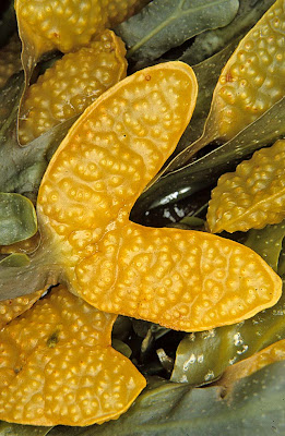
Brown seaweeds in the genus Fucus are common in the intertidal zone. Two species are visible here - saw wrack Fucus serratus with a saw-tooth edge to the fronds and bladder wrack F. vesiculosus with smooth frond edges and paired flotation bladdersIn spring they make rapid new growth and enter their reproductive phase, producing swollen receptacles at the end of the fronds. 


The receptacles are covered in large numbers of small swellings called conceptacles, each of which opens via a minute pore called the ostiole (double click image to enlarge). 


This is a section through a receptacle showing two conceptacles developing inside. This is from a female conceptacle. The radiating, elongated filament-like structures are sterile hairs (paraphyses) and the club-shaped structures are oogonia, each of which produces eight eggs (oospheres)....


...... and here is an egg (oosphere) being liberated from an ostiole into the surrounding water. Inside the conceptacle some oogonia are still dividing by meiosis to produce oospheres - you can see the cell walls forming.


When the conceptacles are mature eggs and vast numbers of swimming male cells (antherozoids) are liberated into the water of the rising tide - most prolifically during spring tides  - and at high tide the eggs are fertilised, if they are lucky, and carried away by the falling tide. If they're luckier still the fertilised zygotes attach to a rock and develop into a new seaweed. The clusters of small bright yellow structures that you can see here amongst the rounded oospheres are the antheridia that produce the antherozoids - this conceptacle is hermaphrodite, showing that it came from spiral wrack Fucus spiralis; saw wrack and bladder wrack have conceptacles that are either male or female.


You can find images of thin sections and male and female conceptacles of fucoid seaweeds here, more detailed information on their structure and life cycle here and more on Fucus and other seaweeds here