Monday, May 24, 2010

Orange-tip Butterflies - More Than Just a Colourful Set of Wings?


This tiny orange rugby ball is the egg of an orange-tip butterfly  Anthocharis cardamines, attached to the flower stalk (pedicel) of a hedge garlic Alliaria petiolata plant in my garden. The caterpillar that hatches will feed on the host plant's developing seed pod, so the caterpillar must hatch soon after the flower is pollinated.


At higher magnification it's possible to see the beautifully patterned egg case, which will be the first thing that the caterpillar will eat when it hatches, before moving on to consumer tender young seed pods.


Female orange-tip butterflies (male seen above with wings open and shut) only lay a single egg per infloresecence, typically on lady's smock Cardamine pratensis but also on hedge garlic A. petiolata or on sweet rocket Hesperis matrionalis, although egg laying has been recorded on 35 members of the cabbage family (Cruciferae). You can see a fine photo of an orange-tip egg on lady's smock over at Stuart Dunlop's Donegal Wildlife blog. If more that one egg is laid per inflorescence the caterpillars resort to cannibalism, but the female butterfly can detect a pheromone signal left by another that has already laid an egg and will avoid that plant, minimising the risk of this gruesome outcome - unless it rains, when the pheromone is washed off. 

Back in 1997 research at Monk’s Wood National Nature Reserve established that the female butterflies that use lady’s smock as a larval food plant are extremely selective in their egg laying habits. They choose large flower heads in open, sunny locations. Choosing a flower head with a large number of buds ensures that there will be enough food (seed pods) for the hungry caterpillar.

Research in Durham University back in 1983 suggested that they also have quite a narrow window of opportunity for egg laying – if the eggs are laid when the inflorescence is too old the developing pods will be too tough for the larvae to eat by the time that they hatch. This can sometimes happen if bad weather delays egg laying, as the butterflies are only active in sunshine.

In my garden this delightful butterfly breeds on all three of the larval food plants - lady's smock, hedge garlic and sweet rocket - which have overlapping flowering periods that collectively span a period of about six weeks. Each plant has different pod development characteristics - hedge garlic, for example, has much larger, tougher, faster developing pods than lady's smock - so I wonder whether any member of the orange-tip population in my local colony can breed on all three - in which case they might need to adjust their egg laying habits to suit the individual host's pod development rates - or whether there are sub-populations that specialise on breeding on each of the different possible hosts. If the latter is the case, that would respresent the first step in subdivision of the population and of one species splitting into three .............. evolution in action...... or maybe they're not that discriminating. Something worth closer study though, I think. With that in mind, I'm planning to breed the butterflies on lady's smock and on sweet rocket, which represent the extremes of the range of flower timing, then see if the resulting butterflies exhibit a preference for laying eggs on either host plant when they hatch.

Sources:
S.P.Courtney and A.E.Duggan (1983) The population biology of the orange tip butterfly Anthocharis cardamines in Britain. Ecological Entomology 8, 271-281.

Dempster, J.P. (1997) The role of larval food resources and adult movement in the population dynamics of the orange-tip butterfly (Anthocharis cardamines). Oecologia 111 (4), 549-556.

Tuesday, May 18, 2010

So Much for Intelligent Design ....


At first glance it might seem that the elaborate mechanism that dandelions Taraxacum officinale use for presenting pollen to visiting insects is a masterpiece of functional design. Look across the top of a dandelion flower with a magnifying glass and you can see a forest of stigmas, divided and curled back at the top of a long style covered in pollen. This is the last stage in a developmental process that begins in the flower bud ....


.... where at this stage the individual florets that make up the flower head (capitulum) are just on the point of flowering. From the bottom upwards in the photo above, first you can see the ovaries that contain the egg cells that will become the embryo in the seeds, then above them are the stamens, joined in a long yellow cylinder.....


... seen here in a single floret. Notice how at this stage the ring of feathery hairs (the pappus), that will carry the mature seed aloft on the breeze, is already well developed. This floret is one from the centre of the flower and has no petal, unlike those around the edge that have ray petals for advertisement ....


... like this one, where you can see the single petal attached. At this later stage of development the style has now elongated inside that cylinder of stamens, forcing its way upwards like a piston and sweeping out the pollen as it goes, then splitting at the tip to reveal the receptive stigma where pollen delivered by a visiting insect will germinate.

The outer surface of the style is covered in a forest of short hairs that help to sweep the pollen out of that cylinder of stamens. Pollen adheres to the outseide of the style until an insect arrives and collects it, at the same time cross-pollinating the stigma with the pollen from another that it arrived with.


But to the dandelions, all of this elaborate floral choreography is redundant - a waste of energy. At some point in their evolution they acquired a mutation that allows their ovules (above) to develop into seeds without any need for pollination, producing clonal, identical copies of the parent plant. It's a process called apomixis, that's also found in some other plants, including some bramble species. So in dandelions all that complex and energetically expensive floral development and the provision of pollen and nectar to attract pollinating insects, now serves no purpose - it's a legacy of an earlier stage in evolution, when dandelions did need to be cross pollinated. In some species of dandelion the pollination mechanism is still functional, but not in the apomitic common dandelion. Perhaps, at some point in the future, mutations will disable the pollen- and nectar-producing mechanisms in apomictic dandelions and they'll be able to shed the cost of producing these expensive resources for no purpose. For the moment, though, all that redundant pollen and nectar is a wonderful resource for bees in spring ....

Monday, May 10, 2010

Four-eyed Fly


These remarkable eyes belong to a male St. Mark's fly Bibio marci - which has not one pair of compound eyes, but two. This is the black fly that dances just above the grass on spring days, dangling its long hind legs. You can find a picture of this behaviour on Nyctalus's Stand and Stare blog. These dancing males are on the lookout for females, which they approach from below, and the conjecture is that those long, fine hairs in between each individual compound eye lens (ommatidium) somehow help in the fly's detection of movement above and precise positioning when he grabs a female.

Remarkably, male and female St.Mark's flies have quite different eyes and are also distinctive in other respects. In this mating pair the larger female is above, with the more slender head, heavier body and smokey-coloured wings.

Both sexes have one pair of exceptionally long legs, that dangle below in flight, although the extent of the difference in leg length is over-emphasised in this picture of a male because the front two pairs of legs have curled up in death.


This head-on view of the male reveals a distinctive horizontal groove across each eye, just below the mid-point. In fact upper and lower eyes are quite separate on each side and have separate connections to the brain, where the images they produce are processed separately - the upper eye on the look out for females, the lower monitoring ground position. This fly has four eyes (as do whirligig beetles which swim on the surface of ponds and simultaneous look towards the sky and down into the water).


The female's heavier body, smokey wings and narrow head are evident in this side view, while ....

... a close-up view of the female's head reveals that her eyes are smaller and hairless - but then she doesn't have to worry about finding males; they find her with their strange two-tier visual system.

Thursday, May 6, 2010

Orchid Roots: Botanical Sponges

You can crudely divide orchids into two groups: ground orchids, rooted in the soil - like Pleione species, for example - and epiphytic orchids like the one below, that often grow on the branches of trees in tropical forests. The dangling roots of the epiphytic types have a dual role, sometimes anchoring the plant and always acting as storage vessels for water that they absorb from mist and sudden tropical downpours. If you cut a section through one of these roots (above) you can see their unique structure, that allows them to absorb and store water. The bright yellow ring of thick-walled cells at the bottom of the image above is the plant's internal plumbing - the pipes (xylem vessels) that conduct water from the roots to the leaves and flowers. Beyond that the thin-walled blue cells are the packing cells that are alive and contain some chloroplasts, like the leaves. Beyond that, sheathing the root and separated by a distinct layer of mostly hexagonal cells is an outer sheath of dead cells called the velamen layer, and their role is to soak up water as the roots dangle in the air, high above the forest floor. They are, in effect, a sponge.

The recommended way to water tropical orchids, like Vanda species for example, is simply to stand them in water for a few minutes each day, so their root velamen layer fills up with water, then simply hang the plants up with their roots dangling in space.

You can see here what happens when you water an orchid root here. When it's dry (above) the dead velamen layer cells reflect light and the whole root looks silvery-white. Make them wet for just a few seconds (below) and those dead cells fill up with water, become translucent and reveal the green photosynthetic tissue within.


Tuesday, April 13, 2010

Potatoes: the Inside Story

These beautiful objects are potato starch grains. Slice a potato, shake the slices in a glass of water and the water will become milky due to the release of starch from the broken cells. Put a drop of this milky water on a microscope slide, viewed it under a microscope using polarised light and this is what you see. The starch itself is colourless and translucent - polarised light is responsible to generating the colours and the distinctive ‘Maltese Cross’ pattern in the grains. Starch grains are polymers of glucose molecules and are the means by which plants store energy for future use. It is breakdown of starch in stems, roots, rhizomes, buds and leaves that is - as this very  moment – releasing the energy that plants are using for new spring growth, before photosynthesis in their leaves takes over the job. It’s also starch that provides most of the calorie intake for almost all of the human population on the planet.
The form and size of starch grains varies depending on plant species and in potato they are relatively large – up to one tenth of a millimetre in diameter. Very fine starch, such as that produced in the tubers of cuckoo pint was used to stiffen cloth and was used by the Eliabethan courtiers to stiffen their magnificent ruffs.

Wednesday, March 31, 2010

Botanical Flypaper

The tiny insect in the photo above, just a couple of millimetres long, is doomed. Its body and wings are held fast by the sticky leaf hairs of.....
.... this plant, a butterwort Pinguicula moranensis that originates from Guatemala and Mexico. Like all butterworts, it captures small insects on its leaf surface and then, when they die of exhaustion, slowly digests them.
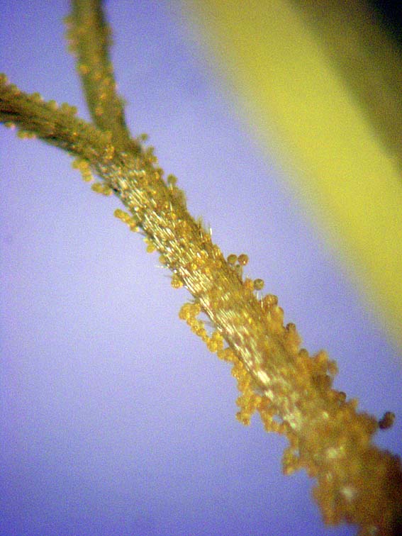
Almost the whole of the plant surface is covered with these minute stalked hairs, of varying heights for maximum trapping efficiency,each tipped with a droplet of sticky mucilage.
Seen here at higher magnification and in side view, each bottle-shaped hair is composed of a single cell rising from one of the surface epidermal cells, topped with a glandular cap that at higher magnification still...
... is revealed to be made up of eight separate secretory cells, each shaped like a slice of cake, perched on the top of the stalk. Meanwhile, down below and embedded in the leaf surface.......
... there's a different kind of gland, seen here in surface view amongst the jigsaw puzzle-shaped epidermal cells of the leaf. Each leaf upper surface is studded with hundreds of these glands. Once and insect is trapped the glands nearby........
..... like this one, seen here in side view at higher magnification, secrete digestive enzymes. When the insect finally dies....
... it collapses into the pool of digestive enzymes and is slowly dissolved, until only its outer chitin exoskeleton remains, like a ghost of the plant's victim. Then the plant absorbs the resultant 'soup', rich in the essential nitrogen that's lacking in this carnivorous plant's boggy habitat. However, not all insects succumb so easily. The plants in my conservatory almost always host...
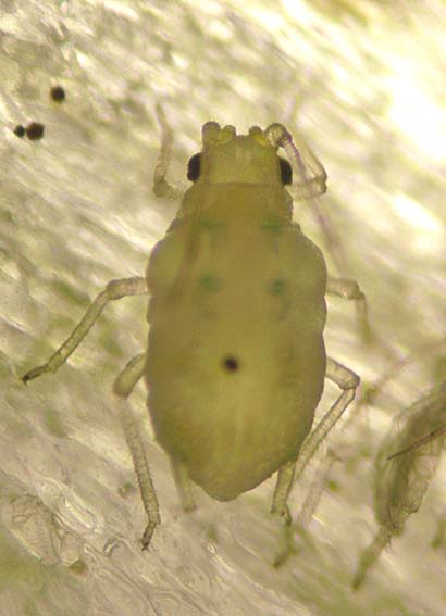
... small colonies of to these tiny aphids. Even though they are held fast, they can still use their piecing mouthparts to puncture the plant's cells and feed, and survive long enough to produce the next generation of young, which are born by virgin birth (parthenogenesis) without the need for mating.  If you double-click on this image for a larger view you'll see a pair of minute claws at the tip of each aphid leg. On most host plants these would allow the aphid to grip the plant surface and walk, but the epidermal cells of butterwort are so smooth and slippery that the claws cannot grip. If you watch under a microscope, you can see the claws simply sliding over the plant surface, so the anchored aphid can do nothing other than feed and breed before it eventually dies, leaving a ghostly shell and a clone of itself behind.


Butterworts' flypaper-like properties make them very useful plants to grow if you are troubled by the tiny mushroom flies that emerge from potting composts - a single plant will trap and kill scores of them.

Sunday, March 28, 2010

Grey Killer

Spring is a rollercoaster ride of hope and despair for gardeners, as tender new seedlings run the gauntlet of frosts, pests and diseases. This fungus, grey mould Botrytis cinerea, is one of the worst killers of plants grown in poorly ventilated, cold clammy greenhouses. Initially, it usually colonies dead or damaged plant tissue like last season's leaves or stems ....

... producing a furry coating for spore clusters on short aerial hyphae.
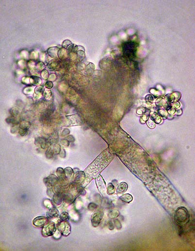
The fungus can produce these clusters of spores, known as conidiospores, in vast numbers, and at higher magnification you can see...
... that each hyphae is branched at the tip. You can also see the cross-walls in the hyphae that indicate that this is an ascomycete fungus
At high magnification the tip of the hypha can be seen to branch, with clusters of spores at the end of every branch....
... that are dispersed on the breeze as a grey cloud when infected plants are disturbed. Grey mould is a major killer of plants but paradoxically it does have its uses. Grapes that are infected with 'noble rot' - as the fungus is known in viticultural circles, produce a much more intense flavour, as the fungus withdraws water from the grape and concentrates the flavour..... a property that's exploited in the production of sauternes dessert wine.

Sunday, March 21, 2010

Anticlockwise tubeworms

The calcareous spiral tubes of tubeworms, attached to wracks and kelps that are washed up on the strandline, are a common sight on the seashore. There are several different species and the first step to identification is to see whether the tube coils clockwise or anticlockwise. If it's clockwise, then it'll be a species of Spirorbis but if it's anticlockwise, like these, and the tube has three distinct ridges, then it's a worm called Janua pagenstecheri. The coiled tube is about 2mm. in diameter.
If you watch the live worm under the microscope it soon everts its crown of transparent feeding tentacles. If you look just to the right of the tentacles you can see a brown, translucent flap. This has a dual function, closing off the tube when the worm withdraws its tentacles and acting as a brood chamber for the worm's embryos. The pink encrustation in front of the worm is a alga, not part of the animal.

Tuesday, March 16, 2010

Fungal Artillary


Most fungi tend to be associated with autumn but there are a number of perennial species that can be found at any time of year, including this one - variously known as King Alfred's cakes, cramp balls or Daldinia concentrica. The first name refers to King Alfred's culinary accident while hiding from marauding Danes in the humble abode of a cowherd; the second refers to the folklore that carrying this fungus around in your pocket stops you getting cramp in the legs (doesn't work for me); the last refers to .....

.... the concentric rings of annual growth that you can see if you cut the fungus open.
The blackened surface of the fungus is covered with scores of these 'pimples', each with a pore in the centre. Each leads to a chamber below, packed with tubular flask-shaped fungal hyphae called asci, each with eight ascospores inside. Cut one of these chambers (in mycological parlance a perithecium) open and this....
... is what you see under the microscope - rows or rugby-ball shaped spores, seen here at around x100 magnification and ....


.... here at x400 magnification. In spring each ascus of eight ascospores elongates in turn, until its tip protrudes from the pore in one of those surface 'pimples', like a cannon protruding from the gun port of a man 'o war. Pressure builds inside the ascus until it ruptures and fires out its salvo of spores. Then it withers, another elongates to take its place and the discharge is repeated. This can go on for 6-7 weeks before all the asci have fired their broadsides, with most of the spore discharge taking place at night. You can watch this by placing the fungus in a light beam in a warm room - if you've got sharp eyes you can see what look like little puffs of smoke all over the surface - the fungus firing its silent broadsides. In England Daldinia concentrica mostly grows on ash trees but in Scotland it also grows on birch.

Sunday, March 7, 2010

Another Living Jewel


My last post showed a jewel-like case made by a single-celled amoeba. This one shows the remarkable case made by a marine worm.  We found this little tapered tube, about 5 cm. long, on the sandy beach at Warkworth in Northumberland this afternoon. It was made by a worm called Pectinaria koreni and when the animal inside is alive only the last few millimetres of the narrow end of the tube protrudes above the sand. The worm lives head-down in the sand, drawing in a current of water through the narrow end of the tube.


You can see the dark zone at the narrow end here - that's the bit that normally protrudes above the sand. The tube is made up of hundreds of sand grains and minute shell fragments, selected for smoothness inside and outside the tube and ....


.... neatly fitted together with a degree of precision that a stonemason would envy....


.... and although the tube is only one sand grain thick it's remarkably strong. That's because....


... the worm secretes a form of cement that glues the grains together, like mortar in a wall ......


.... as you can see here at higher magnification.


A pair would make rather fine ear-rings, provided the wearer didn't have any qualms about wearing jewellery made by a worm rather than by a jeweller.

You can see a picture of the worm here.