Thursday, September 23, 2010

Harvestman

I found this delightful little harvestman amongst some raspberries that I picked yesterday evening. Although they are carnivores, they do seem to have a liking for ripe fruit.
Despite their spider-like appearance and the fact that they belong to the Arachnids, harvestmen are not true spiders but belong to an order of their own known as the Opiliones, distingished from spiders by their globular bodies, unlike those of true spiders which are divided into two parts - the separate thorax and abdomen, separated by a constriction. Harvestmen always seem to have an other-worldly appearance and, scaled up to monstrous proportions, wouldn't be out of place in a science fiction movie. Many of their sensory functions are located in their legs and if you watch the way in which they use these - particularly the second pair that are often far longer than the others - it's quite clear that they are using them to feel and taste their way around their habitat. Nevertheless, when danger threatens they can shed a leg (autotomy) and leave it twitching on the ground, to deflect the attention of  a predator. Unlike true spiders, they can't regrow limbs after moulting, so this desperate measure leaves them short of a limb and sensorily deprived. Before they resort to limb-shedding, they often exude an unpleasant-smelling liquid from their leg joints which you can smell if you hold an irritated harvestman in your hand. This isn't an infallible deterrent - I've watched robins feeding their brood with harvestmen.


One of the most remarkable features of harvestmen is the arrangement of their eyes, in a kind of turret called an ocularium, high up on their back. This arrangement makes sense when you get down to a harvestman's eye-level ...

... when it's clear this this gives it 360 degree vision around and above its globular body....


..... although one can only guess at how much detail those unblinking black eyes resolve.

Nevertheless, whatever angle you approach from a harvestman always has it covered........one of those eyes always seems to have you in its sights...

There's an excellent little booklet called British Harvestmen by J.H.P. Sankey and T.H.Savory (Synopses of the British Fauna (New Series) No. 4  ISBN 0 12 619050) that not only provides fascinating detail about their biology but also contains some delightful little anecdotes. For example, some species apparently kill their prey by positioning their globular bodies over their victim that's imprisoned by their legs and then bouncing up and down on those long legs, 'pile-driving' the unfortunate prey. Others have been noted for a prediliction for marmalade sandwiches from a picnic and on one occasion ink from ink wells (although this one refused black ink and would only drink the red stuff). 

I haven't got around to identifying the species of this individual yet and if there's anyone out there who can help me out, I'd be grateful....

Monday, September 13, 2010

Moth Pointillist Colour Patterns


I found this herald moth Scoliopteryx libatrix, with these beautiful orange markings on its wings, in my garden.


The whole moth is covered with scales, of various shapes, sizes and colours over its whole body, even to the extent that its legs are clothed in this rather fetching pattern of alternating black and white rings.


The main body is covered with fine hair-like scales, but the scales on the wings are....


... much broader, although they vary in width and colour. One interesting feature is that the patches of colour that look fiery orange to the naked eye are composed of a mixture of pinkish-red scales interspersed with variable numbers of yellow scales. The whole effect is reminiscent of colours produced in Pointillist paintings, of the kind made famous by George Seurat. By Seurat's day (1859-1891) the study of colour had revealed that the close juxtaposition of points of two colours could produce the effect of a third colour when viewed from a distance and Seurat exploited this in his meticulously executed paintings. The computer monitor screen that you are viewing this blogpost on uses a similar principle, of coloured dots, to produce its vast range of colours. Butterflies and moths have been exploiting the same phenomenon for millions of years, to either make themselves conspicuous to mates or generate camouflage patterns.
The herald moth's wings also carry small clusters of distinctive white scales, like those just above the 'orange' patch here, which I suspect may be scent scales that emit pheromones recognised by other individuals of the same species.

Monday, September 6, 2010

Brittle Stars

The swaying fronds of red seaweed that fringe rockpools near the low tide level on the seashore are home to a wealth of miniature marine life, less spectacular than the inhabitants of coral reefs but every bit as intriguing. I found scores of these tiny brittle stars, the largest no larger than a centimetre across (including arms), on a visit to the Northumberland coast at the weekend. Brittle stars, or ophiuroids, are relatives of starfish and sea urchins, in the phylum Echinodermata (which means spiny skin - a feature many members of the phylum share). The view above is of the underside of one of the brttle stars, showing the mouth fringed with five teeth formed from calcareous plates.

Seen from above, five arms radiate from the pentangular body. Each arm is formed from articulated segments linked by muscles and these are very flexible, so the animal often curls the tip of an arm around a seaweed frond to stop itself from being washed away by currents. If it's alarmed the muscles between the arm segments contract and then the arms become very brittle.....


... and it doesn't take much force to snap them, as has happened here with the upper arm. This is not a problem, as....

... arms can easily be regenerated, as is happening here with the middle, lower arm. This capacity for shedding and regenerating arms is analogous to the way that lizards shed their tails (autotomy) if they are picked up by that appendage.

At higher magnification you can see the anatomy of the arms more clearly. Each calcareous segment bears spines and a pair of tube feet, that are all interconnected by a hydaulic system of radial canals that run along the arms and a ring canal that runs around the central body. Local relaxation or contraction of muscles, compressing liquid within, elongates or retracts the tube feet.
Unlike the tube feet of starfish which have suckers on their tips and are used for 'walking', those of brittle stars are primarily for sensory purposes and to assist in feeding, by secreting adhesive mucus. In this higher magnification image you can see that the tube foot is hollow.

The ring of tube feet around the mouth on the underside, where the arms converge, help to sweep food particles beyond the five calcareous teeth)....
 

.... into the muscular oesophagous, and then into ....

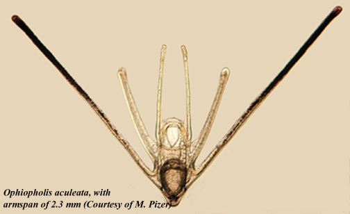
... the stomach. The tiny central body also contains gonads, that produce eggs and sperm that give rise to the planktonic ophiopluteus larvae.

When they're fully grown some brittle star species can reach 60 centimetres in diameter (not in Britain, though), but they all begin life as planktonic larvae, often settling into the shelter of seaweeds on the nursery slopes of rock pools or coastal shallow seas, which are of such importance for the health of the oceans.

You can see a YouTube vieo sequence of an adult brittlestar here.


Thursday, September 2, 2010

Plant Plumbing


Swiss cheese plant Monstera deliciosa is commonly grown as a decorative house plant but in its native Mexican rainforests it's a rampant climber, using its adventitious roots to cling to trees and climbing in much the same manner as ivy in temperate woodlands. Those holes in the Monstera leaf, whose resemblance to holes in Swiss cheese account for its name, let flecks of sunlight filter through to the layers of leaves below, all of which are transpiring water from their surface. If you cut a section through the stem, you can see the internal pipework that conducts water from the roots to the leaves.


In this transverse section of adventitious root, stained with fluorescent dyes that colour dead, woody cell walls yellow and living cellulose cell walls blue you can see the various cells that conduct liquids up and down the root. Embedded in that thick-walled strengthening tissue that gives the root (which in this case is used for clasping tree trunks and branches - this plant is a tropical climber)  rigidity and are fluorescing yellow, are large vessels that conduct water in a continuous tensile column from the roots to the leaf, pulled upwards by evaporation from the leaf surface. The smaller tubes, lined with a layer of blue-fluorescing cell walls, may be resin ducts. The outer cells on the left, part of the ring of small bundles of living cells that encircle the root, are the phloem cells that conduct sugars manufactured by photosynthesis in the leaf to other parts of the plant. Swiss cheese plants are such familiar items of interior decor that they hardly attract a second glance, but they have extraordinary hidden beauty, only visible under the microscope

Friday, August 13, 2010

Defensive Weapons

The outer layer of cells on a plant's surface - the epidermis - is the first line of defence against herbivores, pests and diseases so it's not surprising that many plants are covered with an array of defensive weapons. Sometimes these are cells that secrete repellent biochemicals, which give many plants a characteristic aroma when you brush their leaves. Other species have mechanical barriers, in the form of dense coverings of hairs (trichomes) to deter small insects like aphids. Stinging nettles are covered in a forest of complex stinging hair cells, each mounted on a pediment of cells. You can see some further, more detailed images of the structure of the stinging hairs here, but the image above is an aphid's-eye view of a nettle leaf underside - although they wouldn't see it in these lurid colours, which I generated using polarised light.

Wednesday, August 4, 2010

Gastrotrich

I've been struggling to capture a decent image of a gastrotrich for quite a while, but this afternoon this obliging example (Chaetonotus sp., I think) paused just long enough in my field of view for me to record it. Gastrotrichs are miniature aquatic equivalents of the roadrunner, always on the move - although in this case propelled by a layer of beating cilia on their underside (gastrotrich means 'hairy stomach') rather than legs. This one seemed to be feeding. That circular mouth, surrounded by a ring of tiny teeth, and the muscular pharynx that it leads to ingest just about anything that the animal collides with and that's small enough to enter the gap. The outer covering of cuticular spines give this gastrotrich a fearsome appearence and if you are smaller that it is (i.e. less than about a quarter of a millimetre) the sight of one of these high-speed hunters hurtling towards you must be a nightmare - not that pond life suffers from nightmares; sorry, lapsed into anthropomorphism there. Been watching too many horror movies.  


This specimen came from the edge of a Phragmites reed swarm, amongst the rotting debris in shallow water - a favourite habitat for gastrotrichs. You can see here how agile they are, capable of turning in their own length through 180 degrees. Those two appendages on the tail are adhesive organs. Gastrotrichs do sometimes rest and when they do they glue themselves to something convenient with adhesive secreted from the tips of those appendages. When they are ready to go again they secrete a releasing agent - that, and the glue, which both work under water, must be very interesting substances.

Monday, July 12, 2010

Pond scum


Three weeks of warm weather had left my pond covered with large slimy masses of 'blanket weed' or 'pond scum', the filamentous green algae that tend to plague ponds that have too much nitrogen in the water. When I'd fished most of it out I took a look at a few filaments under the microscope and - like so many living organisms - it revealed structures of great beauty when it was magnified a few hundred times. Inside each cell in the filament the chloroplasts were arranged like strings of green pearls. Various filamentous algae have chloroplasts in different conformations and the most familiar is the spiral chloroplast in Spirogyra..... but this is a different genus.....


The series of fine rings that you can see around the bottom of the upper cell on the left here, just above its junction with the cell below it, identify this alga as a species of Oedogonium. A ring forms each time a cell of this genus divides, so this cell appears to have divided three times.


In amongst the algal filaments there were also desmids - this crescent moon-shaped example is Closterium. The clear areas at the tips of the 'moon' are vacuoles, that contain insoluble crystals of calcium sulphate - a diagnostic feature of this genus.


The most interesting alga in my pond, however, was this one - Coleochaete. It may look like just a pad of simple cells (with some of them apparently dead) but this is an organism of great evolutionary significance. Modern molecular biological studies, and comparative investigations into the ways in which cells divide in this species and in land plants, indicate that Coleochaete shares a common ancestor with present day land plants - mosses, liverworts, ferns, conifers and flowering plants. At some point - maybe half a billion years ago - algae like this, perhaps living in a warm pool of nutrient-rich water like my garden pond, started to colonise the mud and begin the long series of evolutionary changes that led to the development of today's terrestrial vegetation.

A discovery like this makes the chore of cleaning out the garden pond a whole lot more interesting.......... 

Thursday, June 24, 2010

Rolling and Tumbling


This colony of organisms is Synura, a member of the Golden or Chrysophyte algae, that I found in a water sample in the shallows of a reed swamp. Each member of the colony has a pair of unequal length flagellae (to fine to see at this magnification), whose constant beating sends the colony rolling and tumbling through the water. It's about a fifth of a millimetre in diameter. When high concentrations of Synura build up in water bodies they impart a fishy taste, posing a problem for the water industry.

A static image doesn't do justice to it, but the short video clip below gives a better impression of its constant movement. 

Tuesday, June 15, 2010

Another Rotifer....

I've posted pictures of rotifers before (here) but they are such amazing animals that there's always room for another - especially this one, which looks like a monster from a sci-fi horror movie. I found it at the edge of a pond today and it is - I think - a species of Euchlanis, which belongs to a group called loricate rotifers. A lorica is a hard outer shell (the word lorica is Latin for 'body armour') that provides protection and maybe has hydrodynamic properties that aid swimming. The protective function is evident in this photograph (above), where the animal's head is withdrawn into its shield-shaped lorica, which has claw-like points guarding either side of the opening. The animal is about a fifth of a millimetre long.


It may be that the shield-shaped lorica also functions like a wing, generating lift as the animal swims through the water, powered by a tuft of fast-beating cilia at the head end-  which you can see beating in the short video below. When at rest the animal can anchor itself with its tail spines, which open and close like a pair of scissors.

These images were taken with differential interference contrast optics, which generate an apparent three-dimensional image of microscopic objects.

You can find out more about rotifers here.

Monday, May 31, 2010

Ahhhh-tishooooo!


For hay fever sufferers, this can be one of the most miserable times of the year, thanks to grasses and trees that produce vast quantities of wind-borne pollen. Nevertheless, pollen grains are beautiful natural objects. The pollen grains above belong to a tropical plant called cup-and-saucer vine Cobaea scandens, which is pollinated by bats rather than by wind (do bats suffer from hay fever? Probably not...).

I stained the pollen with a fluorescent dye called acridine orange, which binds to the surface of the pollen grain and fluoresces yellow when you shine blue light on it - revealing this exquisite pattern of hexagons and pores (showing green here). Each pollen grain is about one fifth of a millimetre in diameter. Plant genera can often be identified by the distinctive pattern on their surface.

The outer surface of pollen is full of minute pits and chambers that contain proteins that allow a plant to recognise pollen of its own species when it lands on its stigma, and reject foreign pollen. It's these proteins that quite literally get up your nose, trigger an allergenic reaction and set you off sniffing and sneezing.

The outer casing (known as the exine) is made up of a polymer called sporopollenin, which is incredibly resistant to biodegradation - which is why palaeobotanists can recover ancient pollen samples from deep in peat bogs and lake beds and extract and identify pollen samples from plants that grew there tens of thousands of years ago. It's a branch of botany that has given some very useful insights into how plant species distributions have changed during periods of rapid climate change, like the one we are experiencing now: studying the past in this way gives an insight in what is likely to happen to plant species in the future.


Since the exine of pollen is so resilient, it passes through the gut of insects unharmed, although the pollen contents are digested. Yes, that little white speck on this bumblebee's tail is bee-poo, made up of empty exines of pollen that it has eaten. Many hoverflies feed almost exclusive on pollen, leaving little piles of hoverfly poo on leaves, and I know of at least one enterprising entomologist who has collected and analysed this, in order to study hoverfly's pollen diet.

During the Vietnam war Yellow Rain - yellow specks coating plants in the jungle - was believed to be the result of Communist chemical warfare. Subsequent anaysis showed that it was bee faeces, produced by vast swarms of bees that sometimes rose into the air and defecated in unison.