Thursday, April 5, 2012

Pond Population Explosion


A combination of unusually warm spring weather and spawning frogs that stirred up the mud and so released a lot of of nutrients into the water recently led to an algal bloom in our garden pond. Large patches of algae floated on the surface in mucilaginous mats that trapped bubbles of oxygen.


Under the microscope, at x40 magnification, the algal cells were round, highly motile and present in vast numbers. This group represents the population in about 2% of a single drop of water.


The same, but at x100 .... and at ....


.... x400. I am not certain what species this is but I think it may be Chlamydomonas. The paired flagella of each cell are not resolvable with this microscope at this magnification.


The most striking aspect of this algal bloom, apart from the sheer numbers of cells, is the hyperactivity of the algae. The bubbles of scum on the pond surface may seem static, but at this magnification they represent a surface film of frantic activity.


Friday, March 30, 2012

Within Every Grass Leaf There Are Hidden Smiley Faces .....


A

A vascular bundle in a transverse section of a grass leaf, stained with the fluorochromes Calcofluor M2R (blue fluorescence = cellulose) and auramine O (yellow fluorescence = lignified cell walls). The red fluorescence is chlorophyll autofluorescing red in the blue excitation beam of the microscope. 

The two big 'eyes' in this 'smiley face' (which is typical of a monocot vascular bundle) are metaxylem elements that transport water through the leaf. The bright blue fluorescence in the 'mouth' of the 'smiley face' is phloem, composed of larger sieve tubes and smaller rectangular (in cross section) companion cells, which together transport sugars, made by photosynthesis, out of the leaf. The bright yellow cells forming the neck of the 'smiley face' are lignified, providing a measure of rigidity in the leaf,  and the band of cells along the bottom of the section are epidermal cells covered by a cuticle.

Friday, February 24, 2012

Seaweed Sexual Reproduction: a chancy business


This is a transverse section of the conceptacle of a brown seaweed Fucus sp., commonly known as saw wrack. It has been stained with a fluorescent dye called anilino-naphthalene-sulphonic acid. 


Brown seaweeds in the genus Fucus are common in the intertidal zone. Two species are visible here - saw wrack Fucus serratus with a saw-tooth edge to the fronds and bladder wrack F. vesiculosus with smooth frond edges and paired flotation bladdersIn spring they make rapid new growth and enter their reproductive phase, producing swollen receptacles at the end of the fronds. 


The receptacles are covered in large numbers of small swellings called conceptacles, each of which opens via a minute pore called the ostiole (double click image to enlarge). 


This is a section through a receptacle showing two conceptacles developing inside. This is from a female conceptacle. The radiating, elongated filament-like structures are sterile hairs (paraphyses) and the club-shaped structures are oogonia, each of which produces eight eggs (oospheres)....


...... and here is an egg (oosphere) being liberated from an ostiole into the surrounding water. Inside the conceptacle some oogonia are still dividing by meiosis to produce oospheres - you can see the cell walls forming.


When the conceptacles are mature eggs and vast numbers of swimming male cells (antherozoids) are liberated into the water of the rising tide - most prolifically during spring tides  - and at high tide the eggs are fertilised, if they are lucky, and carried away by the falling tide. If they're luckier still the fertilised zygotes attach to a rock and develop into a new seaweed. The clusters of small bright yellow structures that you can see here amongst the rounded oospheres are the antheridia that produce the antherozoids - this conceptacle is hermaphrodite, showing that it came from spiral wrack Fucus spiralis; saw wrack and bladder wrack have conceptacles that are either male or female.


You can find images of thin sections and male and female conceptacles of fucoid seaweeds here, more detailed information on their structure and life cycle here and more on Fucus and other seaweeds here

Wednesday, January 11, 2012

Better than Cannabis.....


There was a time – before the advent of synthetic fibres based on plastics and petrochemicals - when the wealth and security of nations depended on tough, coarse plant fibres that provided rigging for sailing ships and the raw material for countless other essential objects  - like sacks and sails for example - so explorers were always on the look-out for new supplies of this strategic material.

 Joseph Banks, travelling on Captain Cook’s first voyage to the South Seas in 1769, had high hopes that he might make a fortune from growing New Zealand flax Phormium tenax that he found in those Antipodean  islands, as a substitute for cannabis fibre which, up until then, had provided most of the fibre for rigging naval vessels. Maoris made their traditional textiles from the Phormium fibres but Banks envisaged a thriving industrial market for the product, whose fibres are much stronger than those of cannabis, and an attempt was made to use convicts to grow the plant as a fibre crop on Norfolk Island. Banks was destined to be disappointed - you can read an account here - but it did become an important source of fibre for rigging in the 19th. century..

The image above shows a transverse section of a New Zealand flax leaf, using the fluorescent dye auramine O to stain the lignified fibres, which show up as the transverse yellow-green bands in the image. I've turned the natural orientation of the leaf 90 degrees clockwise, to fit the page.

The thick-walled fibres have a tiny central cavity (the lumen), which is typical of sclerenchymatous fibres. 

The transverse red bands are photosynthetic parenchymatous cells - chlorophyll fluoresces red in the blue light that was used to illuminate the specimen.

The bright blue cells are bundles of thin-walled phloem, which has no lignin in its walls, and the brighter yellow cells surrounding the phloem will be lignified xylem, conducting water.

The lower surface of the leaf, to the left of the image, has a lignified hypodermis, below the epidermis.

The natural function of the fibres and lignified hypodermis is to provide structural rigidity for the long, narrow, sword-shaped leaves, which are held upright in the living plant, which is illustrated below (public domain image from Wikipedia Commons http://upload.wikimedia.org/wikipedia/commons/a/a5/NZflaxPiha02.jpg


Today Phormium tenax is mostly grown as a decorative garden plant.

Friday, December 30, 2011

Micro-rock-pooling in Winter.


It's too cold in winter to spend a lot of time paddling around in rock pools but you can always take a few samples of seaweed home on a jar of seawater and have a look at the smaller inhabitants under the microscope. These two, each about a millimetre long, were in a  sample of Corallina officinalis seaweed. The upper specimen is an unusually bristly acarine mite, found clambering through the seaweed fronds. You can see more acarine mites by clicking here.

This is a minute flatworm, with two very simple eyes, found gliding over the surface of the seaweed, propelled by thousands of cilia that are only visible at high magnification under the microscope. You can see another marine flatworm, in more detail and with a movie of the cilia in action, by clicking here.

Friday, December 23, 2011

Sowing Wild Oats


Some seeds need to be sown while others - like wild oat Avena fatua - sow themselves. This is a wild oat fruit (or, to be botanically accurate a caryopsis) in the dry state. It's equipped with a long awn (which is extension of the floret in which  the fruit formed and in which it is shed), that's bent at a right angle about a quarter of the way along it's length.


When the caryopsis falls to the ground and gets wet - from a passing shower of rain, for example, that bent awn straightens, then bends again as it dries out. The picture above shows the same fruit, but now it's been moistened and the awn has straightened. As the awn bends and straightens it also rotates, because the awn is constructed from a helix of fibres that twist and generate torsion as they dry (see below).


The outer coat of the floret containing the caryopsis is equipped with this arrowhead of stiff hairs at the tip ....


.....which readily catch in fur and feathers and help disperse the seed, but also anchor it in crevices in the soil when it falls to earth.


There is also a beard of stiff hairs running up the groove in the caryopsis. As the awn rotates .....


.... with the expansion and contraction of this helical tube of spiral fibres that it's constructed from, it levers the caryopsis further into soil crevices. Those stiff hairs on the caryopsis help to anchor it in the soil, ratcheting it in ever deeper until it's in a  moist enough position to germinate and put down roots. 

This is a seed that sows itself.

The video below shows a group of wild oat caryopses writhing as their awns dry out and begin to rotate.


Sunday, November 27, 2011

Breathing Space


This is a thin section of the lower stem of water milfoil Myriophyllum sp. , stained with the fluorochrome calcofluor which binds to the cellulose of the cell walls and is fluorescing brightly in ultraviolet light. Marsh plants tend to be rooted in anaerobic mud and so have air channels (aerenchyma) that conduct oxygen down to the roots. 


Working from the outside inwards in this section, there is a well defined single outer layer of very small cells forming the epidermis, then inside that lies the stem cortex with 17 air channels arranged around the central stele, which contains the phloem (brightest flourescence) and the xylem.


The stem and leaves of water milfoil. The small white structures in the leaf axils are the stigmas of the female flowers.

Thursday, November 10, 2011

Plant Cuticles


The surface of plants (with a few exceptions, such as those that live submerged under water) is covered with a tough, transparent, waxy layer called the cuticle, composed of cutin secreted by the layer of epidermal cells that it covers. The best way to see the cuticle is to snap the leaf of a drought-adapted succulent plant like Crassula ovata and pull one part of the leaf against the other, peeling away the cuticle, which covers the above ground parts like a wrapping of cling-film.


These  cuticular peels, often with a single layer of epidermal cells attached, can be mounted on a microscope slide .....


..... and viewed to reveal the pattern of cells and the .....


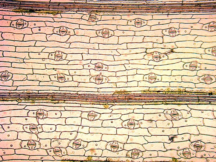
...... stomata, which allow carbon dioxide to enter for photosynthesis. The two images above are of cuticular peels of spiderwort Tradescantia virginiana.


In this vertical section of a leaf, stained with fluorescent dyes, the cuticle appears as the bright yellow layer on top of the epidermal cells. In the centre you can see a single stoma in vertical section, with closed guard cells and its adjacent subsidiary cells, with a sub-stomatal cavity below leading to the loosely packed mesophyll cells, where photosynthesis takes place. The cuticle has a dual function - keeping water in and keeping the leaf surface dry. 


In plants adapted to arid conditions (xerophytes), like this Aloe variegata  , the cuticle is visible as an extremely thick transparent layer that allows very little water to escape from the leaf.


In plants that are subjected to frequent rainfall, like this nasturtium Tropaeolum majus leaf, fine grooves in the cuticle surface trap air below the water droplets, which then round-up under their own surface tension and simply roll off the leaf when it shakes in the wind. This is vital, as a wet leaf surface blocks stomata and prevents carbon dioxide from entering, slowing down photosynthesis. As the water rolls off the leaf it carries away dirt and dust, so the leaf cuticle is effectively a self-cleaning surface - a phenomenon known as the 'lotus effect', referring to the extremely hydrophobic self-cleaning leaves of sacred lotus. The silvery area under the central water droplet in the image above is caused by minute air bubbles, trapped between the water and the leaf surface.


The cuticle can also have a secondary defensive role, as seen in this painfully prickly leaf of the hedgehog holly Ilex aquifolium 'ferox', which is covered in cuticular spines. In general, leaves of evergreens, that survive for several years before they are shed, tend to have thick cuticles that protect the leaf against herbivore attack throughout their extended life.


Thursday, November 3, 2011

Springtails


Every time I lift the lid of our garden compost bin scores of these tiny insects, each smaller than the diameter of the head of a pin, leap around in all directions. They are members of the ancient insect order known as the Collembola - commonly called springtails - and feed on decaying vegetation. The darkness, warmth and humidity of the compost bin suits them perfectly. I think this species might be Folsomia candida, which is very common in gardens.


Most of the time they move slowly on those stumpy legs but when they are alarmed they hurl themselves into the air using an organ called a furcula under their tail .....

.... which you can see in this specimen. You could liken its action to a kind of exceptionally energetic pole-vaulting. The tip of the furcula is held in place by a clip-like structure called a retinaculum, but when the muscles in the furcula contract the clip suddenly releases its grip and the furcula flicks downwards and backwards, hurling the animal upwards and forwards.

You can find pictures of another springtail species here and a fine set of photographs for ID purposes here.

Monday, October 31, 2011

Was Sid, the Mystery Microscopist, a Scotsman?


When I was exploring the old microscope slides belonging to the mysterious 'Sid' that I mentioned in my previous post I came across one that was clearly home-made and was labelled as porphyritic olivine basalt from the Lion's Haunch, Arthur's Seat, Edinburgh. The hill is the plug from the mouth of a long extinct volcano, exposed after softer sedimentary rocks were eroded from around the igneous core.  Polarised light microscopy reveals the crystalline inclusions very nicely.

Similar rock samples were collected on the Apollo 15 mission to the moon in 1971

Arthur's Seat is the very conspicuous hill - some might call it a mountain - almost in the centre of Edinburgh. From the right angle it looks like a resting lion and this polished thin section of igneous rock came from the 'Lion's Haunch'. Of couse, there's no way of telling whether Sid, who was apparently a chemist, prepared this specimen himself or swapped it with a geologist friend - microscope slide exchange clubs were once common - but he clearly had an interest in Scottish geology, so may have been a Scotsman......